By: Mai Sas
PhD candidate Mai Sas, working with Dr. Phil Shane, has flown over to Japan in 2018 to analyze some of her samples using a CAMECA 1280-HR multi-collector secondary ion mass spectrometer (MC-SIMS), a high-resolution instrument not currently present in New Zealand. Mai is looking at oxygen isotopic compositions of quartz crystals from rhyolitic deposits from the Okataina Volcanic Centre, a dormant caldera system in the Taupo Volcanic Zone of North Island, New Zealand. Mai is trying to understand if, and how, oxygen isotopic compositions fluctuate at the Okataina magmatic systems, and what implications that may have for this, and other, rhyolitic volcanic centres. She completed her analyses at the Isotope Imaging Laboratory (IIL), which is part of the Creative Research Institution (CRIS) at Hokkaido University in Sapporo, Japan, an excellent facility with several SIMS instruments. The images below follow include a glimpse at the initial sample preparation stages, as well as which analytical instruments were used at IIL.
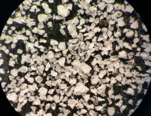
Microscopic (low-magnitifcation) image of the crushed pumice sample, showing a mixture of minerals and volcanic glass. The majority of the minerals consist of plagioclase and quartz, with some ferromagnesian minerals such as biotite, Fe-Ti oxides, and orthopyroxene. Each mineral had to be examined carefully to make sure it is the desired mineral, as plagioclase, quartz and fresh volcani glass can all appear very similar. Helpful identification tools include crystal form and mineral cleavage or fracture.
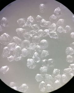
Microscopic (low-magnitifcation, but higher than previous) image of the picked quartz crystals from one of the tephra samples.
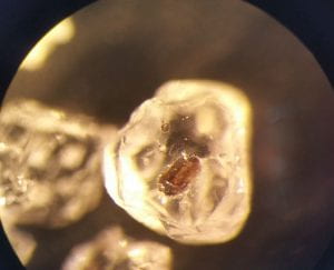
Microscopic (higher-magnitifcation) image of a picked quartz crystal with an inclusion.
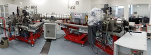
CAMECA 1280-HR MC-SIMS. The SIMS instrument takes up the space of a large room and is situated in a clean lab, with full body suits and air blasts required prior to lab entry. This ensures quality analysis and prevents contamination.
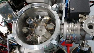
CAMECA 1280-HR MC-SIMS sample dock, where the samples sit under vacuum prior to analysis.
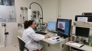
CAMECA 1280-HR MC-SIMS working station, with the SIMS located in the room with the glass windows.
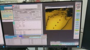
CAMECA 1280-HR MC-SIMS high-spatial resolution analysis of a quartz crystal. Spot size is ~20 microns in diameter.
Mai examining each SIMS spot post-analysis to ensure no inclusions were tapped and that fracturing of the sample didn’t occur. This was completed via high-magnification back-scatter electon (BSE) imaging using a JEOL JSM-7000F field-emission scanning electron microscope (FE-SEM).
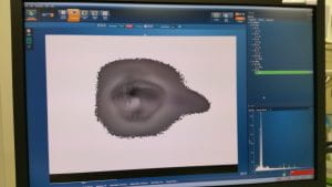
BSE image of a SIMS pit in quartz. Pit is ~20 microns in diameter.
